
Retina Blog

Unveiling the sub-ILM haemorrhages
January 25, 2026
This video presents a series of non-resolving pre-retinal haemorrhages involving the macula that required surgical intervention. Clinically, distinguishing subhyaloid from sub-ILM haemorrhages can be challenging. Intraoperatively, high-magnification visualization helps identify the exact plane, guiding ILM peeling when the bleed is sub-ILM. The video highlights these magnified views to differentiate the planes of pre-retinal haemorrhage.
1.4 MM Trophy
January 21, 2026
A 47-year-old man presented after a metallic particle struck his left eye while using a power saw. VA hand motion; IOP 8 mmHg; RAPD, ciliary injection, 2-mm hypopyon, 2+ cells. B-scan: dense hemovitreous; CT: large metallic IOFB on inferior retina. Dense hemovitreous revealed an inferior IOFB with RRD. An inferior L-sclerotomy allowed 0.12 forceps removal (enlarged, then closed). Endolaser, fluid–air exchange, PFCL removal, 1000-cSt silicone oil, and methotrexate used. Retina remains attached.
Better to be safe than sorry
January 15, 2026
33 Y.O patient with a total RRD in one eye without PVD, multiple peripheral breaks and macular involvement, and extensive predisposing lattice degeneration in the fellow eye. The affected eye was managed with combined encircling scleral buckle, phacoemulsification with IOL implantation, and 23-gauge pars plana vitrectomy using PFCL, endolaser, and C3F8 gas tamponade. A prophylactic scleral buckle was later placed in the fellow eye. Bilateral anatomic success achieved and maintained for both eyes

FLANGED IOL IMPLANTATION
December 23, 2025
This video demonstrates the technique of flanged intraocular lens (IOL) implantation in cases of surgical aphakia, particularly in eyes with large corneal diameters (>12 mm), where conventional scleral-fixated IOL methods carry a higher risk of postoperative IOL slippage or excessive tilt. The flanged approach provides stable fixation, improved centration, and enhanced long-term IOL stability in challenging anatomical situations.THE CRIMSON TIDE
December 22, 2025
Crimson Tide showcases vitrectomy in advanced proliferative diabetic retinopathy complicated by dense fibrovascular membranes, asteroid hyalosis, and severe intraoperative bleeding. The video highlights the use of tissue plasminogen activator assisted large clot removal over a detached retina thereby helping in salvaging the eye and preventing post operative membrane formation under silicone oil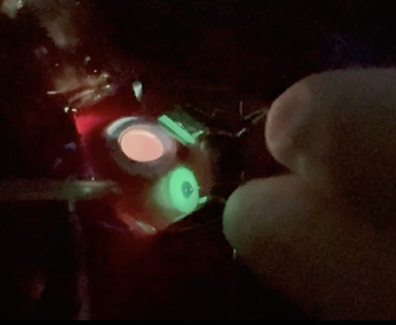
THE GLOWPORT
December 16, 2025
The Glowport is an innovative illuminated vitrectomy trocar cannula designed to enhance intra-operative visualisation and streamline instrument handling during vitreoretinal surgery.Findings suggest that the Glowport significantly enhances port visibility, reduces intraoperative errors, and may improve overall surgical efficiency. Its affordable, reusable design makes it a valuable tool for both seasoned surgeons and trainees.
VITREOMACULAR TRACTION ASSOCIATED WITH MACULAR HOLE.
December 15, 2025
Vitreomacular traction (VMT) occurs due to incomplete PVD with abnormally strong adherence to macula. A 68 year old male had decrease vision in RE. On examination, he was found to have VMT with macular hole as evident on OCT. Patient was taken up for surgery. Posterior hyaloid should be removed in a circumscribed manner to avoid any undue traction over foveal edge. Inverted ILM flaps along with gas tamponade resulted in successful closure of macular hole.
POWER OF THE NEEDLE.
December 15, 2025
Repair of combined retinal detachments in diabetics is very challenging. Bimanual dissection of fibrovascular membranes with silicon oil injection is required to achieve good anatomical and functional outcome. In this video, a needle has been used along with Max grip forceps for bimanual dissection. The membrane was lifted with forceps and needle was used to separate the attachments of fibrovascular membrane and underlying retina. It reduces the incidence of iatrogenic breaks in retina.- « Previous Page
- 1
- 2
- 3
- 4
- 5
- 6
- …
- 26
- Next Page »