
Retina Blog


The “Not-so-Foreign” Body
September 30, 2026
17-year-old male with open-globe trauma and suspected metallic IOFB presented with corneal tear, dense vitritis and HM+ vision. Underwent corneal repair, 25G PPV, lensectomy, IOFB removal, intravitreal antibiotics/steroid and silicone oil. Surprisingly, the “metallic IOFB” was TWO EYELASHES. At 1 month, SOR with secondary sulcus IOL was performed. Vision improved to 6/12.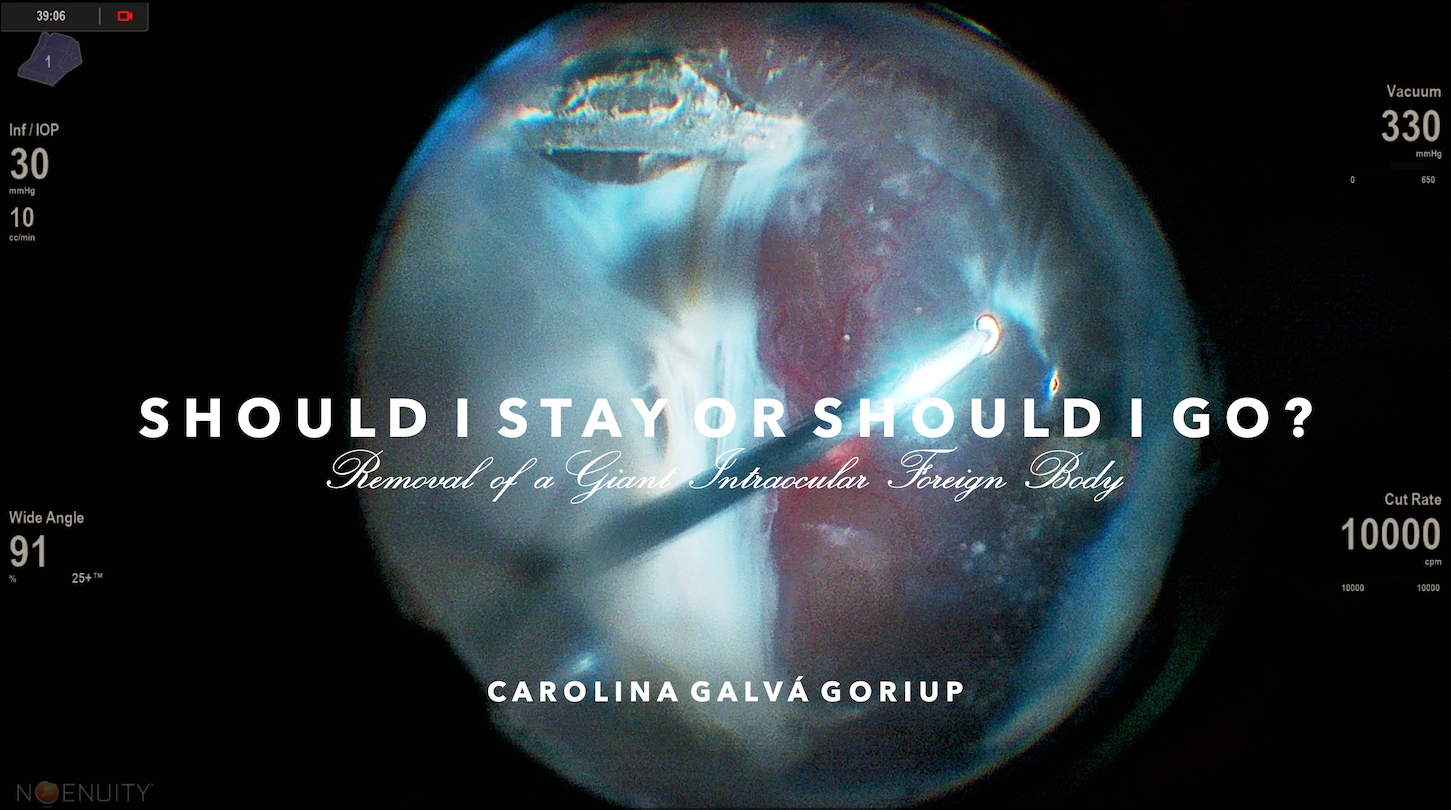
Should I Stay or Should I Go — Removal of a Giant Intraocular Foreign Body
September 23, 2026
This is the case of a 37-year-old male who presented one month after penetrating ocular trauma with a large metallic IOFB embedded in the inferotemporal retina and associated retinal detachment. After lens aspiration and vitrectomy, heavy liquid and cohesive viscoelastic were used to protect the retina during extraction. A T-shaped scleral incision was performed, and a Castroviejo 0.12-mm forceps was used to remove the 7-mm metallic foreign body.
Intracapsular Cataract Extraction of Sublimed Black Crystalline Lens
September 16, 2026
In this video, the surgeon trims vitreous around a sublimed crystalline lens to allow it to prolapse into the vitreous. The lens is then attempted to be removed via fragmatome but given the density is unable to be broken down. The surgeon proceeds by using a scleral tunnel based intracapsular cataract extraction technique and uses two vials of PFO to float the lens into the anterior chamber and out of the scleral tunnel wound.
Recurrent retinal detachment and macular hole repair with AmnioGraft
September 15, 2026
76 year-old male with history of macula off retinal detachment in the right eye complicated by recurrent superior detachment and new full-thickness macular hole 6w post-op from their initial vitrectomy. Full-thickness macular hole repaired with AmnioGraft. Pre-operative and post-operative fundus photos and OCTs included.

THE TRIPLE THREAT
August 13, 2026
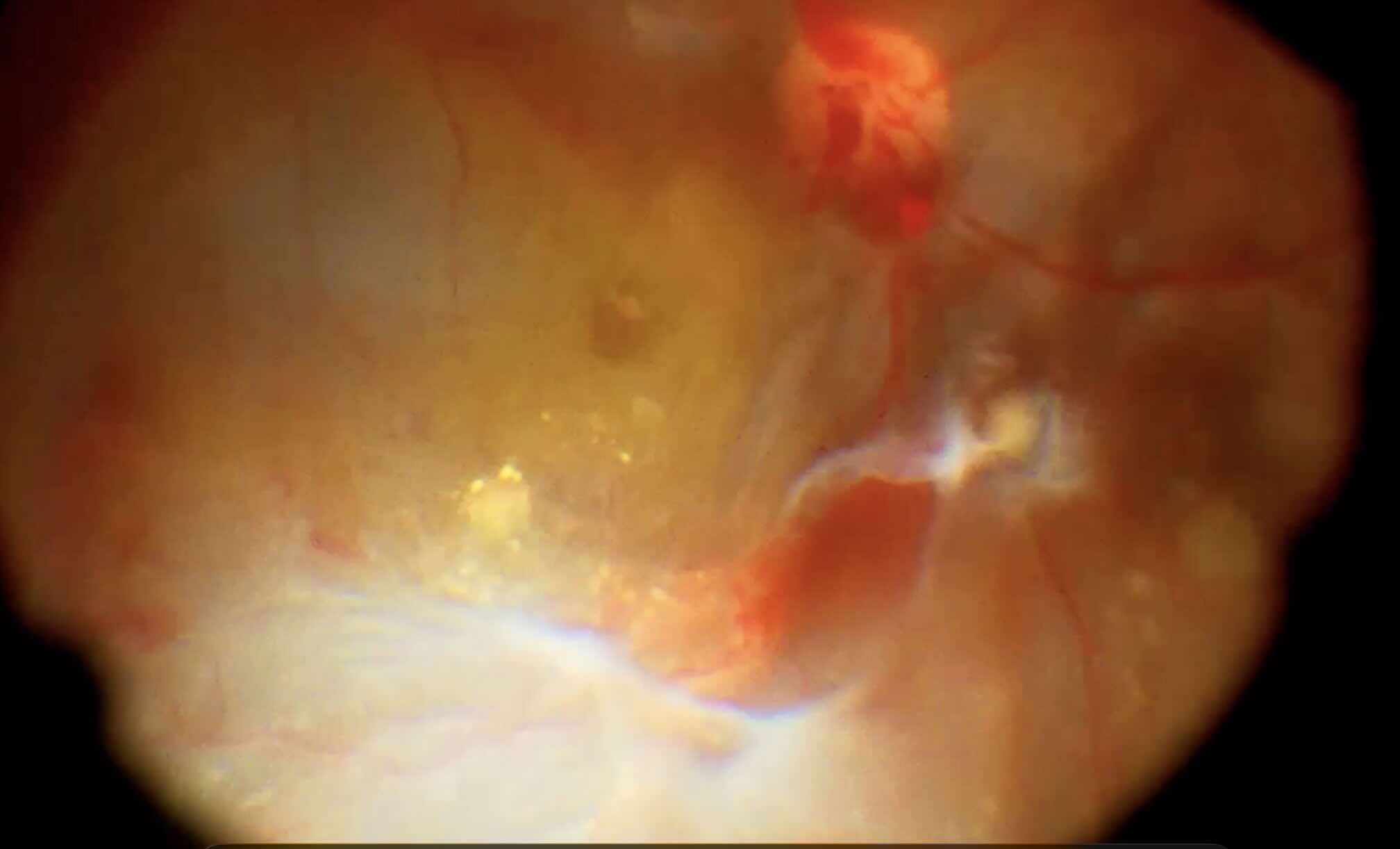
Surgical video demonstrating pars plana vitrectomy for a combined retinal detachment with a full-thickness macular hole. Following meticulous vitrectomy and vitreous base shaving, selected peripheral tractional membranes were preserved to avoid iatrogenic retinal breaks. The macular hole was repaired using a temporal inverted ILM flap, with silicone oil tamponade. Following oil removal, the retina remained attached with successful hole closure and good visual recovery.- 1
- 2
- 3
- …
- 25
- Next Page »