
Retina Blog
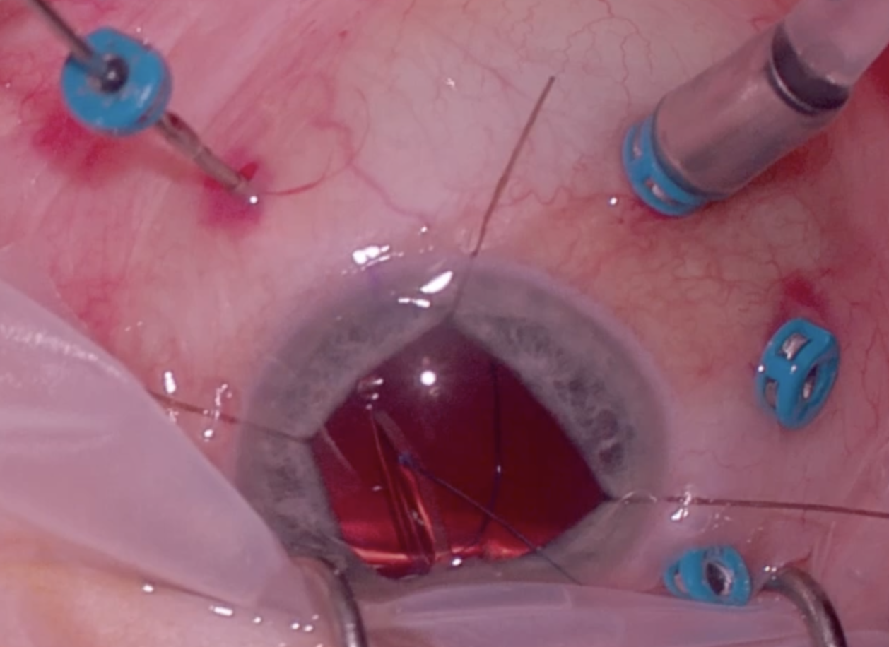
Trocar Assisted Yamane Secondary IOL
June 17, 2026
This is a video of removal of dislocated intraocular lens (IOL) with placement of secondary IOL using the trocar-assisted Yamane technique.
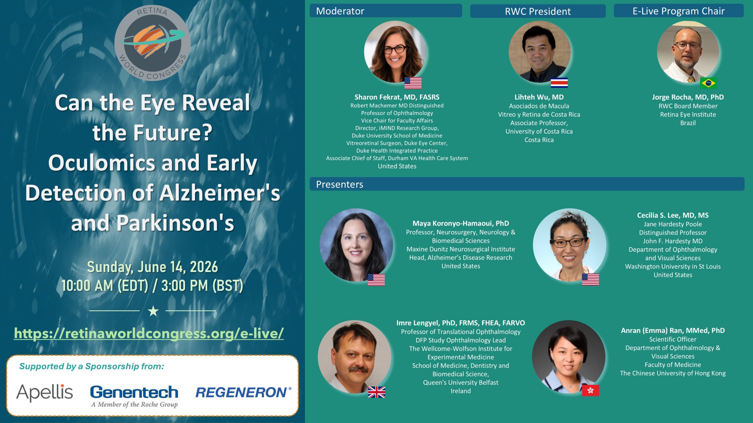
ELive 060726 Can the Eye Reveal the Future? Oculomics and Early Detection of Alzheimer’s and Parkinson’s
June 7, 2026
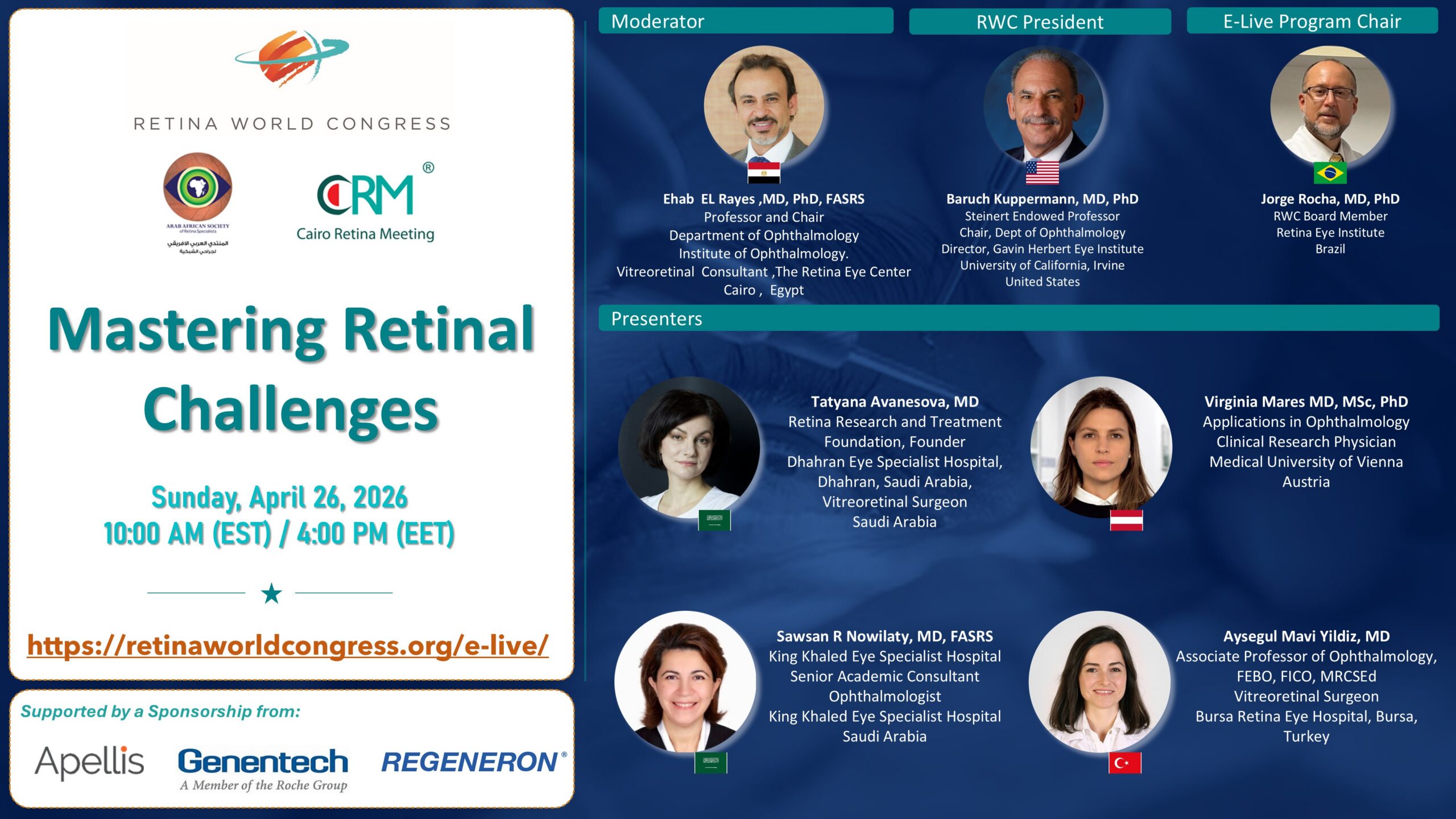
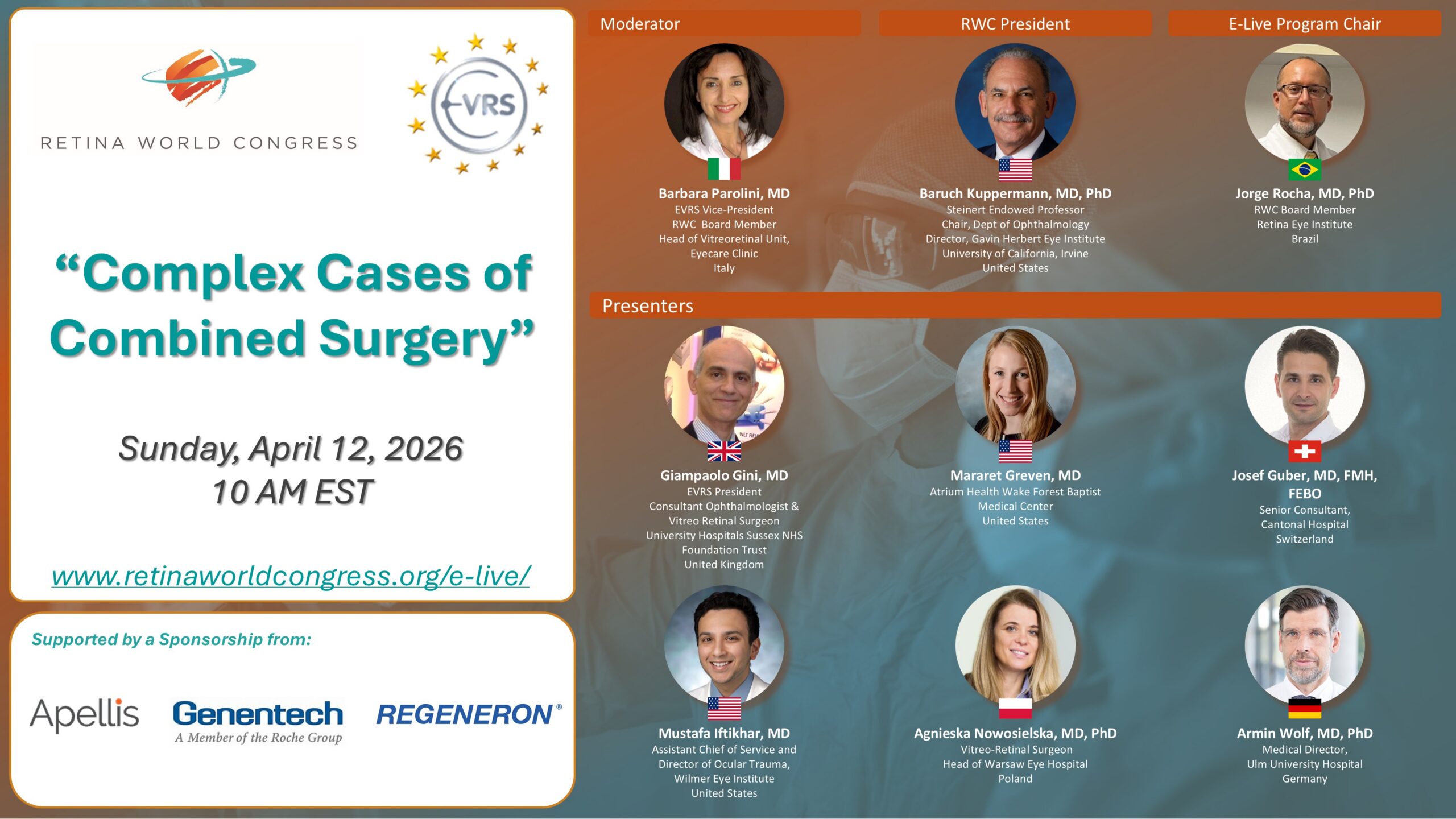

Management of 180° Iridodialysis with Aphakia and Total Hyphema
March 20, 2026
A complex case of abandoned SICS cataract surgery presenting with aphakia, total hyphema, and 180° inferior iridodialysis. The video demonstrates intraoperative findings and surgical repair of the iridodialysis to restore iris configuration.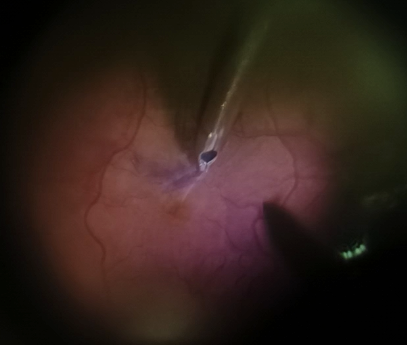
The Thin Line Beetwen Foveal Sparing Peel in Abnormal Adhered ERM Cases and Macular Hole Creation
March 8, 2026
This is a case of a usual moderate epiretinal membrane in the left eye of a 64 year-old female, pseudophakic patient. BCVA was 20/60. During pars plana vitrectomy, abnormal adherence was noticed during ERM peel. This way, in order to avoid macular hole creation, we decided to discontinue complete ERM peel, sparing the fovea while trimming the redundant ERM membrane. ILM peel was extended in order to minimize ERM recurrence.

Repair of Closed Funnel Retinal Detachment Through Boston KPro
February 22, 2026
This patient initially presented with a severe keratitis which led to a corneal perforation that required multiple transplants. Ultimately, a Boston KPro was placed. Upon presentation to our clinic she had a dense retroprosthetic membrane and a closed funnel retinal detachment. In this video we show the repair which was a challenge. Ultimately, we were able to reattach the retina after complex and extensive tissue manipulation.
- « Previous Page
- 1
- 2
- 3
- 4
- 5
- …
- 26
- Next Page »